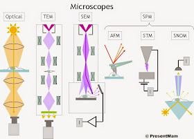
Micoscopy
The microscope is the first powerful tool used in biological
studies
Microscope is the instrument to magnify minute object , there
are many different version of microscope present today era are
1. Bright
light Microscope :-
Most common microscope used today’s biologist
It having two lenses system, The ocular lens (eye
piece) & object lens
It visible light range is between 4000-7000 Å wavelength
It able to magnify upto about a thousand times of
actual size
The resolution power of the eye is about 0.1mm , while
that of microscope is 200mm
The resolving power of the microscope is determine by Abbe
Equation
2. Dark Field
microscope :-
Commonly
used for achieving a marked contrast between living organisms and the
background dark field microscopy
In these sample is viewed only with oblique ray , it
is particularly used for studying suspension of bacteria
In these person sees those rays that are scattered
from objects, it highlights specimens against dark background
3. Phase
Contrast Microscope :-
Zernicke (1932)
introduced the phase of principle contrast microscopy
He was awarded By Nobel Prize for that discovery
These microscope enhances the contrast between cells
& their environment & between internal organelles & their
surroundings
It is commonly used for observation living or
unstained cells
Person can see mitochondria, mitotic division ,
nucleoli & other organelle quite clearly than other in these
4. Interferance
Microscope :-
Like phase
contrast microscope , it functioning depend on functioning in the speed of the
light passes through different materials.
It has wider applications, it is relatively easy to
vary contrast between & colour effect
It is mainly used for quantitative determinations,
such for obtain dry weight
It has highly accurate optical balance, unlike phase
microscope
5. Polarizing
Microscope :-
It is mainly used to viewing highly ordered objects
such as crystal or bundles of parallel filaments
The mitotic spindle formation can be studied by these
microscope
6. Ultraviolet
microscope :-
It uses shorter
UV rays for shorter wavelength (1500-3500 Å)
It lens composed of fused quartz , calcium fluoride or
lithium carbonate
It is used for Qualitative & quantitative determination
of nucleoplasm
7. Cofocal
scanning Light Microscope :-
In late
1950s Marvin Minsky invented revolutionary new instrument, which is produce an
image of a thin plane situated within much thicker specimen
The Specimen is illustrated by a finely focused laser bean scans across
the specimen at a single depth
8. Flourescence
Microscope :-
It uses UV
light with higher wavelength stimulation on cell components
It is most often used to detect specific proteins
& other molecule in cells & tissue
In these a very powerful & widely used technique
used is to combine fluorescent dyes to antibody molecules
In these two common fluorescent dyes are used
Flourescein (Gives green) & Rhodamine (Gives red)
 |
| Types Of Microscopes |
9. Electron
Microscope :-
M.Kholl & E.Ruska (1932) invented the electron
microscope.
It is ideal instrument for studying cellular infrastructure
There are two major electron microscope are employed
are Transmission electron microscope & Scanning electron microscope
Transmission electron microscope is most common used
in these electron microscope
Scanning electron microscope especially useful for
examining the surface of specimens
10. Scanning Tunnelling Microscope :-
It is
invented by two scientists Gerd Bening & Heinrich Rohrer (1981)
It produces bumps &valleys of the atoms on the
surface of object
Mapping of surface done by “Feel” much as blind person
explores the surface of ground by tappng a cane
Gerd Bening & Heinrich Rohrer got Nobel prize (1986)
for inventing Tunelling micoscope
No comments:
Post a Comment