Sunday 29 June 2014

Structure of Megasporangium


Structure of Megasporangium (Microsporophyll = Carpel) :-

also called Ovule 

=>  Structure Of carpel Differentiated Into 3 Part

i) Upper Recepetive Part = Stigma
ii) Elongated Tube Like Structure = Style
iii) Lower Swallon part = Ovary
These Are Arrange above the thalamus

@ Structure Of Megasporangium :-


Nucellus :-


 Protect Inner Part especially Embryo sac surrounded by two integument . Which present inner side called inner integument & Which present outerside called outer integument
* Portion of embryo where egg found called micropyle . Which help in entry pollen tube
* Back to micropyle end , there is broad part called Chalazal end
* Funicles is the part which help in Attachment of ovule from placenta

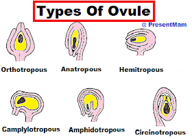
@ Types of Ovule :-


1. Orthotropous :-

Straight type ovule , in this ovule micropylar & Chalazal end found opposite to each other in straight line
- These type of ovule have Devoid of curvature

2. Anatropous :-

These type of ovule is completely inverted having 180 degree curvature of funicles
- In these type of ovule micropyle found near to hilum or faces toward placenta

3. Hemitropous :-

In this ovule Degree of curvature between orthotrophous & Anatropous , when body found right angle to funicles

4. Camplylotropous :-

In these type , ovule became somewhat curved so micropyle & chalazal come near to funicle , In these Nucellus became slightly curved & funicle attached to the middle of body of ovule

5. Amphidropous :-

in these type , nucellus & embryo sac both become curved & converted into horse-shoe shaped 

6. Circinotropous :-

these type ovule is orthotrophous in young condition , but due to elongation of funicle .it beome slightly inverted & funicle cover the total ovule because unilateral growth of funicle

Saturday 28 June 2014

Development Of Male Gametophyte


Development Of Male Gametophyte :-

Male Gametophyte development Happen in 2 main Step 

1st = Pre-pollination :- 

All the changes or development happens before pollination
* In 70% Dicot, Microspore Divide into 2 Parts
* In Rest 30% Dicot, Microspore Divide into 3 Cell Before Pollination

NOTE :- 


When Microspore Start germination found Inside Microsporangium Called Precoceous

2nd = Post-Pollination :-

All the Changes or events Happens After & During pollination

@ Microspore :- 


is a firstly developed male gametophyte

@ Character :-


 It have Dense cytoplasm , lot's of lubish body & large vacoule


@ Development :- 

- Vacoule Became Large pushes nucleus toward bottom side of microspore
- Nucleus Divide by unequal Division , Cells attach to wall form lense shape
- After Some time Cellulose Cementing substance Deposit outside germinating cell get apart & Independent cell form generative cell
- Now, This cell called Pollen grain (Which are Partially germinated)
- Now Pollen Grain released at in 2 cell or may be 3 cell from anther
- After That Released 2 or 3 celled pollen grain react to outer surface of stigma by agency of water, wind or animal etc called Pollination
- Development of male gametophyte on stigma Involve steps are :-
i) Absorption of Water by pollen grain
ii) Swelling of pollen grain with in few minute
iii) Releasing of Wall recognition Factor

NOTE :-


 These Factor Decide Wheather pollen grain germinate or not on that particular stigma
iv) After Mutual Recognition Tube cell enlarge & intine Come out in form of pollen grain after rupturing Exine
v) Pollen tube grow within cell of stigma in transmitting tissue of style
vi) Pollen tube secrete pactenase & Hydrolytic Enzyme , These enzyme produce entry of pollen tube also absorb nourishment
vii) Tube cell & germinate cell move at tip of pollen tube with regular cellulose deposite in the tube
viii) Germinate Cell Divide Form male gamete
ix ) All back part became Non-functional , due to deposition of of cellulose
x) Tube cell degenerate before fertilization, they function as give nourishment to sperm cell

Friday 27 June 2014

Types of microspore


Types of microspore :-

i) Tetrad antherii) Isobilateral antheriii) Linear tetrad antheriv ) T-shaped antherv) Decussate anthervi) Compound anther :-


Found In asdepiadacae Family , Microspore of an ather lobe remain united with each other & form sac like structure pollirium

@ Microsporangium Structure :-


They are haploid , Minute , Uninucleated, Brown Structure found in many number inside microsporangium formation after meiosis of spore mother Cell
- Outer most layer Called Sporoderm Consist Exine & Intine

- Exine :-

it is outermost layer composed by Sporopollenin ( Organic Chemical polymer which is resistant from oxidation leaching ) Ubish Body

- Intine :-

These Intine consist Pectocellulose
- In Angisperm Exine form From tapetum during Sporogenesis
- In angiosperms Intine form from development of male gametophyte

@ Exine Part :-

i) Sexine
i.i) Endoexine
i.ii) Tactum
ii) Nexine
ii.i) Endexine
ii.ii) Foot layer

@ Palynology :-

Shape, Size Of microspore & Pollen grain. Exine mIcrosculpturing & Number position Character Of aperture Are great taxonomic importance , These are study under separate Branch Called Polynology
- Exine Surface Of Pollen Grain Having Various Pattern of Microsculpturing & Ornamentation Or Discontinues One or more Placed Exine Is Thin Called aperture or Intactate Area through . which pollen tube emerge out at time of germination on stigma

Wednesday 25 June 2014

Stamen


Stamen :-

They are Modified micro-sporophyll. which are concerned to form microspore

@ Types :-


i) Basified :-

When anther attach to base of anther

ii) Adnate :-

when filament all of spore

iii) Dorsified :-

When filament attach to dorsal side of anther

iv) Varsataline :-

When filament attach to dorssal surface but with very weak point & always move as air flow

@ Development Of Micro-sporangium :-

- In Young Condition at Time of initiation , some part of anther Consist outer Epidermis & inner hypodermis
- After Some time later , just below epidermis some cell become meristamatic & These cell Called archesporangium or archesporial Cell
- During These For transporting of Food & water inner dark ring are formed in middle centre
- Archesporial Cell Form 2 Cell Layer , outer Primary Pariental cell or wall & Inner Primary Sporaneous Cell


- Primary Parietal Cell Divide Form 4 Layer,
* Epidermis
* Endothecium
* Middle Layer
* Tapetum
- After That Inside tapetum layer Microsporogenesis (In Which 2n Mother Cell Divide Into 4 Haploid Microspore) Happen
- And At Last During Dehisense Few Endothecium Cell Break To Form Stronium & Microspore released in air

@ Short Note On Tapetum :-

Inner wall of Microsporangium Wall , consist of single layer wall filled with dense cytoplasm may be uni or binucleated , During Microsporogensis tapetum provide harmone , Enzyme , amino acid & Other nutritive to the sporogenesis mother Cell , In Angiospore Tapetum is of two type :-

i) Gladular Or Secretory Tapetum :-

The Cell Secrete some stem cell During Meiosis Produce proubish Body Within Cytoplasm Coated With Sporopollenin , These Body called ubish Body, Causes thickning Of Enzyme of microspore

ii) Periplamodial or Amoeboidal Tapetum :-

These Cell Dissolve Early by dissolution of wall so The tapetum Protoplast produce multinucleated plasmodium provide nourishment to dividing Microspore Mother Cell

Monday 23 June 2014

Flower is modified Stem


Proof That flower is modified Stem :-

Ans :- 


many , evidence , have been given to proof flower is modified stem are 
i) Position of flower bud & shoot bud is same in plant body , both are terminal & axillary in position
ii) In some plant flower bud modified into vegetative bud , ex :- Onion, Agave
iii) In some plant thalamuss elongate to form vegetative branch of another flower above the first flower Ex :- Rose 
iv) In Lymphia (water lily) flower show all transitional stage b/w sepal to petal & petal to stamen
v) In Cynandropsis , Thalamus elongate show internode b/w petal & Stamen . the internode b/w petal & stamen called androphore & internode stamen & carpel called gynophore
vi) in case of rose sepal having similar to morphology to leaf
vii) In degeneria stamen are expended like leaf, carpel appear like folded leaf with differenting into stigma & style
viii) Anatomy of thalamus , pedicel , stem show close similarity in case of vascular supply & other thing, floral part also show some vascular supply like ordinary leaf

Function Of Flower Part


 Function Of Flower Part 

1. Calyx :-

- It Protect floral Part In Bud Condition
- It Is Green In Colour & It Prevent Rapid Transpiration of Young flower
- Sometime it Change In Various Colour Or Got Petaloid Condition , Which help in Attract insect so directly help in pollination

2. Corolla :-

- Attract insect for pollination by its colour for pollination
- having Fragnance So Help In pollination

3. Androecium = Male Part :-

- Indivisual part of these Called Stamen
- It Produce male gamete
- It Act Main Role In Formation of male gamete & Fertilization
- It Consist Of Three Part :-
* Anther :- Where Pollen Grain Formed & mature
* Connective :- Connective between Filament & Anther
* filament :- Long Tube like structure

4. Gynoecium :- Female Female Part :-

- Indivisual part Of These called Carpel/pistil
- It Consist of Three Part :-
* Stigma :- Receptive Part , In Which pollen grain Bind
* Style :- Long Tube Like Structure , Which Connect Stigma to ovary
* Ovary :- Bulb Like Structure , Where Ovule are present 

@ Some general Terms :-

- Polyandrous :-

 In These Condition Stamen are Free or separated from each other

- Aldalphous Condition


 = These Are Denaoted As Stamen arrangement , which May be many type as :-

* Monoaldalphous :-

 In These Filament Of Stamen Fused to form one group EX= Hibiscous Flower

* Dialdalphous :- 


In These Filament of stamen Fused In Two Group EX= Pea (Having 9+1 Arrangement)

* PolyAldalphous :- 


In These Filament of stamen fused To Form many group EX= Citrus Fruit

- stamenal Tube :- 


These Is a tube which produce by Fusion of many filament of many stamen
- Perianth condition :- 

These are Those Condition In Which Not Distinguish B/w Sepal & Petal of flower , so collectively called Tepal

* When All Tepal Are free Than These Called Polyphyllus Condition

* When all Tepal Are fused than these called gamephyllous Condition


- Monotheca Condition :- 


These is denoted that condition in which all pollen grain fused To form one pollen sac chamber In which pollen grain present

- Ditheca Condition :-


 These Is denoted that Condition In Which All pollen grain form two pollen sac Chamber in which pollen grain present
- Fruit = Is called Fertilized Ovary
- Seed = Fertilized Ovule


- Reniform Anther :-


 When anther Is in Kidney Shape

- Hypogynous Condition :-


 If all part of flower along with ovary present above thallamus/torus

- Perigynous Condition :- 


If all part of flower present above thallamus & Ovary somewhat Present below Thalamus Or embedded on Thallamus

- Epigynous Condition :- 

If all Part of flower present above thallamus & Ovary is Fully embedded in thallamus


@ Syndrous condition :-

Anther & Filament both are fused through out their whole length, found in cucurbitaceous family Ex :- luffa 

@ Syngenecious Condition :-

anther of all filament unit & form a cylinder around style, found in composity family Ex :- sunflower

Flower

 Flower 

@ Flower :-

When Vegetative Branch Of Angiosperms Convert In reproductive Structure called flower

@ Pedicel :-

Stalk of flower
# If Pedicel present Than Called Pedicilate Condition

@ Pediuncle :-

# Stalk Of infloresence

@ Bract :-

At Basr Of Pedicel Small structure present 

# Condition :-

- If present Then Called Bratate Condition
- If Not Present Called Ebractate Condition

@ Bracteoles :-

Leaf like Structure On Mid Of Bracteoles

@ Thallus / Torus :-

braod Structure At Base Of Flower , which hold All Flower Structure

@ Zygomorphic Condition :-

These Is That condition In which flower have bilateral Symmetry Structure

@ Actinomorphic Condition :-

In These Condition Flower have Radial Symmetry

@ Part's :-

- Non Essential Parts :-

these are those Parts of flower in which are not so much importance in flower but help in attracting Insect for their pollination
Ex = Calyx (it's Indivisual part called Sepal) & Corolla ( it's Indivisual part called Petal )

- Essential Parts :-


These are those apts of flower , which have great importance in flower
Ex = Androcium (stamen - male part) & Gynoceium (carpel- Female Part)
Note :-
~ Sometime sepal Get Colour like petal structure so called Petaloid Condition
~ Sometime Petal get Colour like Structure So called Sepaloid Condition

@ Condition :-

* Fused Sepal called GamoSepalous Condition
* If sepal not fused remain individual called Polysepalous Condition
* Fused Petal called Gamopetalous Condition
* If Petal not fused remain individual called Polypeatlous Condition 

@ Pistil / Carpel Consists of :-

- Stigma = receptive in nature
- Style = Tube Help in transport of pollen grain to ovary
- Ovary = Bulb like part , where ovule present & Fusion of gamete occur

@ Stamen consists of :-

- Bilobed anther = Where pollen grain present
- Connective = which help anther to attach filament
- Filament = long thread like structure

Friday 20 June 2014

CHARACTERISTIC FEATURE OF GYMNOSPERMS

CHARACTERISTIC FEATURE OF GYMNOSPERMS:-
# include 70 living genera, 725 species out of 16 genera and 53 species from India.Acc. To Raizad sahni in 1960
# living gymnosperm found in temperate region, cold climate, dry region even artic region# CYCAD'S:-Few gymnosperm found in plain
# gymnosperm are perrenial (live long) ,xerophytic (found on dry climate or water) ,evergreen, aborescent
EXTERNAL FEATURE:-
# plant body is sporophytic differentiate into root, stem and leaf
# tallest gymnosperm = Siquia
Smallest gymnosperm = Zamea
# root system is tap root system
# special feature:- root is associated with algae in symbiotic relationship
# few root also associated with fungi
# stem may me branched or unbranded
# on stem leaf is Scarl are found# leaf may be
1) Monomorphic 2) Dimorphic/ \Folliose. Scale
INTERNAL FEATURE:-
# vascular bundle found in ring
# secondary growth found
# annual ring is found

Thursday 19 June 2014

Angiosperms & Character

Angiosperms & Character :-

- Angiosperms are also called angio-spermatophyta or Anthophyta
- Their Seed present in fruit
- Male & Female gametophyte present inside flower
- These plant are found everywhere in earth
- There leaf venation may have parellel or reticulate type
- In These xylem fiber (Libriform fibers) Present
- True Vessel are present in these plant
- Their stamen may have bilateral symmetry, Having bilobed pollen grain
- In These proper exine are formed
- Zoophily type pollination are shown by these plant ,
- These plant having Colourful petal, fragnance smell, sticky pollen grain
- Highly receptive stigma present
- No Archegonial chamber present
- Their ovule are covered by double
layer integument
- These plant show double fertilization, which results in triple fusion
- These plant have highly reduced female gametophyte
- These plant ovule are 7-celled & 8 Nucleate gametophyte

# Paleobotany :-

In these we study Recovery & identification of plant remain
- Acc. To paleobotanical , angiosperms came in jurassic period & Cretaceous period period & As temperature changes many of modification occur in angiosperms, so it show many of modification & diversification
# Theory About Origin Ogf Angiosperms :-

1. In Paleozoic Period :-

- Here 10,000 old plant laphius seed are found in artic & antartic region
- Many of pollen found in Carboniferous rock
Note :- In these era only micro fossil found not macro fossil

2. In Pre-cretaceous Era :-

- Cornet (1986), Sanmiguella Lewissi firstly found macros fossil of angiosperms
- Secondary, Another angiosperm fossil of Archaenthus Linnenbergeri are found
- This Acc. to Sun et al. discovered oldest angiosperms reported Are Archaefrust.

3. In Lower -cretaceous era :-

in these era there is evidence of development of wood,fruit, leaf & Venation many modification seems
- In These Era only diversification of plant are seen

4. In upper Cretaceous era :-

In These era , their is evidence of angiosperms presence in earth along with fern & Gymnosperm

Tuesday 17 June 2014

List Of Some Quantitative identification of Disaccheride, Polysaccheride & Protein

List Of Some Quantitative identification of Disaccheride, Polysaccheride & ProteinTest For Disaccheride :-

1. Hydrolysis Test :-

- Take 2.5 ml of Sample (Sucrose) in Beaker- Add 4-5 ml drop Of Concentrated HCl into it- Boil Solution Mix it Constantly Till Quantity reduced to half (1/2) - Now Divide Solution into 2 Test tube , Then mark both test tube 'A' & 'B' for Fehling & Benedict Test respectively 

2. Fearjen's Test :-

Colour Test For Polysaccheride :-

1. Iodine Test For Starch :-

- Take 2 ml of Test solution (starch) in Test Tube - Boil & cool it at Room temperature - Then add 2-3 ml of iodine solution

Observation :-

Blue Colour Appear 

Result :-

Presence Of starch is Confirmed

2. Iodine Test For Glycogen :-

- Take 2 ml of Test solution in Test Tube - Boil & cool it at Room temperature - Then add 2-3 ml of iodine solution

Observation :-

Colour Disappear on heating

Result :-

Presence Of glycogen is Confirmed

Test For Albumin (protein)

1. Biurate test :-

- take 3 ml of test Solution in test tube (protein = Albumin) - Add 1 ml of 40 % NaOH- Add 2 drop of 1% of CuSO4

Observation :-

Violet & Pink Colour , Which may dissappear on Adding excess of CuSO4

Result :-

Presence Of protein (albumin) is Confirmed

2. Xanthoproteic Test :-

- Take 2 ml of test solution - Add 0.5 ml of HNO3 into it- Slowly heat it Boil & Cool it under Tap water- Add 40% NaOH to make it Alkaline

Observation :-

Yellow & Turn Orange On Addition Of NaOH

Result :-

Presence Of albumin Is Confirmed

3. Mercury Chloride Test :-

- Take 2 ml of test solution - Add Mercury Chloride Into It 

Observation :- 

white ppt formed

Result :-

Presence Of albumin Is Confirmed

@ Reducing & Non-reducing Test :-

1. Methylene Blue Test :-

- take 3 ml of Distilled water - Add 1 Drop of methylene Blue - Add 1 ml of test solution- Then 1 ml of 40% NaOH

Observation :-

Blue colour Dissappear 

Result :-

Test Solution Contain Reducing Sugar

2. Picric Acid Test :-

- Take 3ml of Test Solution - Add 1 ml of picric Acid Solution into it- Add 1 ml of 40% NaOH solution

Observation :-

Red Colour Appear

Result :-

Test Solution Contain Reducing Sugar

3. Tommer's Test :-

- Take 2ml of 0.5 ml of CuSO4 - Add 2 ml of Test Solution - Add 2 ml of 40% Of NaOH

Observation :-

Yellow Or Brick red Colour

Result :-

Test solution Contain Reducing Sugar

Monday 16 June 2014

Quantitative identification of Simple Sugar

List Of Some Quantitative identification of Simple Sugar (monosaccheride)

@ General Test :-
1. Mollisch test :-
Step are
- Take 2 test tube , mark Then A & B
- Then take 2 to 3 ml of Given sample (sugar) in 'A' test tube
- Then Add 2 drop of mollisch Reagent into it
- Shake test tube 'A' Very well
- Take 3-4 ml Concentrated H2SO4 in test tube 'B' ,
- Then with care , gently add H2SO4 in test tube 'A' from 'B'
Observation :-
While adding H2SO4 into Test tube 'B' , A pink or violet colour ring appear at junction of both while mixing
Inferance :-
given sample contain carbohydrates
@ Test For Fructose :-
2. Faugler's Test :-
Step are
- Take about 5 ml ogf given sample (fructose) in test tube &
- Add 3 ml of faugler sample into it
- Boil & Shake test Tube Gently
Observation :-
Blue colour appear
Inferance :-
Given sample contain Fructose
3. Sawniroff test :-
- Take 3ml of sawrinoff agent
- Add 1ml of test solution
- Boil it for 30 Sec
Observation :-
Red to orange colour appear
Inferance :-
Presence Of fructose is Confirmed
@ Test For Glucose :-
1. Benedict Test :-
- Take 5 ml of benedict Reagnent in Test Tube
- Add .5ml of test solution (glucose) in it
- Mix both solution well & Boil For two minute
- Then cool it under tap water
Observation :-
yellow cream Or Brick red ppt Formed
Inferance :-
Test Solution Contain Reducing Monosaccheride (glucose)
2. Fehling Test :-
- Take 3 ml of 1% potassium ferring cyanide in Test Tube
- Add 1 ml of NaOH solution into it
- Boil & Then Add Near 10 drop of test solution (glucose) & keep it to cool
Observation :-
Yellow colour ppt. of ferricyanide become to fade & finally decolourized
Brick red to Colorless
Inferance :-
Test solution Contain Reducing Ferricyanide

Sunday 15 June 2014

Respiration , Breathing & it's Regulation


PARTIAL PRESSURE :-

of a gas defined , the pressure it exerts in a mixture of gas & calculated as total pressure of mixture of gasseous >< percentage of gas in a micture 

Condition for RESPIRATORY SURFACE :- 

For Cutanious Surface :-

1. It Should be Thin , About 1mm 
2. It Should be permeable for gasseous
3. It Should be moist with water & Mucous always
4. it should be have large surface area
5. It Should be highly vascular (rich blood supply )
6. It Should have direct or indirect contact with source of oxygen
7. Presence with respiratory pigment
8. It sHould have surfactant , very important , which prevent collasping of alveoli

EXCHANGE :-

Pulmonary gas Exchange :-

# Inspired air :-


N2 = 78.6 % ---- 597 mm Hg (Partial Pressure)
O2 = 20.84 % ---- 159 mm Hg (Partial Pressure)
Co2 = 0.04 % ---- 0.3 mm Hg (Partial Pressure)

# Alveolar Air :-

N2 = 74.09 % ---- 569 mm Hg (Partial Pressure)
O2 = 13.6 % ---- 104 mm Hg (Partial Pressure)
Co2 = 5.3 % ---- 40 mm Hg (Partial Pressure)

Transport Of CO2:-

i) As In Form of carbonic acid =(7% of total)
ii)As In Form of Bicarbonate of Na+ & K+ = 70%
iii) As In Form of carboamine haemoglobin =(23% of total)
H2O + CO2 <-------Carbonic anhydrase-----> H2CO3 --- Breakdown ----> H+ & HCO3-

# As Carbonic Anhydrase haemoglobin:-

Hb(-) + H(+) <-----> HHb (Haemoglobinic acid ) reduced form of Hb
HHb + CO2 <----> CO2HHb (carboamine haemoglobin

# calculated Amount :-

3ml/100 ml of bloof
* Atrial Blood = 48ml Of CO2 /ml of blood, which divided into two part
i) Plasma (36 ml)
ii) R.B.C (12 ml)
* Venous Blood - 52 ml of CO2 /ml Of Blood
i) In plasma = 36ml
ii) In R.B.C = 14 ml

NOTE :- 

10 ml of blood carry 3.7 ml of CO2

# Haldane Effect On CO2 Transport 

1. Decrease Binding Of CO2 with Hb , so favour dissociation of Carboamini Hb
2. More Acidic Oxyhaemoglobin , it stimulate release Of H+ ion From Reduced (HHb) , So Increases Formation of carbonic acid 

Transport Of Gaseous :-

1. Transport Of Oxygen :-

These occur in two main step as
i) In Solution form (1-3%) in which only 0.3 ml/100 ml of blood transported
ii) In Oxyaemoglobin (90-97%)
For Calculated Amount = Solubility Coefficient of O2 At 37 degree Celcius & 760mmHg >< Partial Pressure 
= 0.00003ml >< 100 
For actual Amount = 19.80ml/100ml of arterial Blood 
=> Human Body Required 375 litre of oxygen /day , which mean we have to take 1900 litre of fresh air to fulfil demand of 375 litre of oxygen/day
=> 15 gm of haemoglobin /100ml of blood can transport 19.80ml (round off 20ml) of oxygen called 20 volume percentage

@ Requirement Of different Animal Of oxygen :-

Fishes = 9 ml
Amphibian = 12 ml
Reptiles = 9 ml
Birds = 18.5 ml

@ oXygen Dissociation Curve :-


# Significance Of Haemoglobin

- It increases carring capacity in form of oxyhaemoglobin
- it also increases CO2 carring capacity in form of carbonic acid
- also act as acid base buffer
- Readily combine with high partial pressure of O2 & release easily in low partial pressure of O2 into tissue

@ Bohr Effect :-

it based upon Partial Pressure Of CO2 . Which Relates Also with pH, temperature,Oxygen Dissociation Curve move Right side with increase in Hb For O2 resulting Less % saturation

Control Of Breathing :-

Breathing are controlled by some centre in brain named as:-

i) Inspiration Centre :- in medulla oblongata at ventral side in position
ii) expiration Centre :- in medulla oblongata at dorsal side of brain
iii) Peripheral chemoreceptor , which having 2 parts
- aortic arch
- Carotid Sinus

iv) Chemoreceptor In Brain
v) Mechano-receptor or stretch receptor in lung alveoli in branchional tree